Our Focus
The van Rheenen group studies the identity, behaviour, and fate of cells that drive tumour initiation, progression, metastasis and the development of therapy resistance. These populations of cells are difficult to study since they are rare, and their behaviour (e.g. migration) and traits (e.g. stemness) change over time. To be able to study these dangerous cells, we have developed microscopy techniques to visualize individual cells in real-time in living animals, referred to as intravital microscopy. For example, we developed small imaging windows that can be surgically implanted in mice giving visual access to tissues with cellular precision for several weeks. We combine the latest genetic tumour models with intravital imaging to obtain fundamental knowledge on cancer. Our research focuses on three areas are
(1) The cellular mechanisms of tissue development and homeostasis, tumour initiation, and tumour progression; (2) The cellular mechanisms of migration and metastasis of cancer;
(3) The molecular and cellular mechanisms of therapy resistance and side effects.
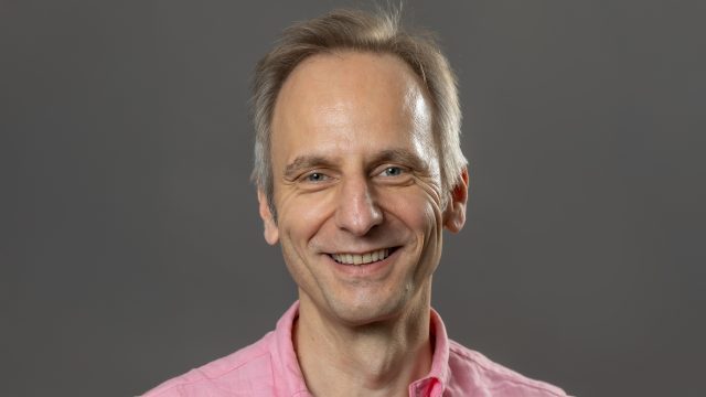
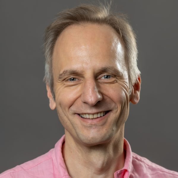
About Jacco van Rheenen

Jacco van Rheenen
My Research
Jacco van Rheenen was originally trained in a variety of imaging techniques during his PhD with Dr. Kees Jalink at the Netherlands Cancer Institute. He was among the first to optimize imaging and develop software to quantitatively measure FRET on confocal microscopes. In order to broader his scales, he obtained a KWF fellowship to do a postdoc in the United States in the lab of Dr. John Condeelis at the Albert Einstein College of Medicine. There he extended his imaging experience by imaging mammary tumors intravitally including two-photon microscopy and became an expert in the field of intravital FRET imaging.
In 2008 he was appointed as group leader at the Hubrecht Institute, where he utilizes his imaging techniques to visualize processes that are required for the metastasis of tumor cells in living animals. In July 2014 he was appointed professor in Intravital Microscopy at the University Medical Center Utrecht. In October 2017 he became senior group leader at the Netherlands Cancer Institute (NKI) in Amsterdam. In 2009, he was awarded a VIDI award from Netherlands Organization for Scientific Research. In 2013, he received the Stem Cells Young Investigator Award. In 2015, he was awarded an ERC consolidator grant, and in 2017 the Dr. Josef Steiner Cancer Research Foundation Award.
Awards
2022 VICI award, Netherlands Organisation for Scientific Research (NWO), Netherlands
2019 Ammodo Science Award, Netherlands
2017: Dr. Josef Steiner Cancer Research Foundation Award
2015: ERC consolidator grant by the European Research Counsel
2013: Stem Cells Young Investigator Award 2013
2008: VIDI grant by the Netherlands Organisation for Scientific Research (NWO)
2006: Fellowship for fundamental and (pre-)clinical cancer research from the Dutch Cancer Society (KWF).
Key Publications
Azkanaz M, Corominas-Murtra B, Ellenbroek SIJ, Bruens L, Webb AT, Laskaris D, … & van Rheenen J. Retrograde movements determine effective stem cell numbers in the intestine. Nature. 2022 Jul;607(7919):548-554. DOI: 10.1038/s41586-022-04962-0
Hannezo, E., Scheele, C. L., Moad, M., Drogo, N., Heer, R., Sampogna, R. V., ... & van Rheenen. J, Simons, B. D. (2017). A unifying theory of branching morphogenesis. Cell, 171(1), 242-255.
Scheele, C. L., Hannezo, E., Muraro, M. J., Zomer, A., Langedijk, N. S., Van Oudenaarden, A., ... & Van Rheenen, J. (2017). Identity and dynamics of mammary stem cells during branching morphogenesis. Nature, 542(7641), 313.
Zomer, A., Maynard, C., Verweij, F. J., Kamermans, A., Schäfer, R., Beerling, E., ... & van Rheenen, J. (2015). In vivo imaging reveals extracellular vesicle-mediated phenocopying of metastatic behavior. Cell, 161(5), 1046-1057.
Ritsma, L., Ellenbroek, S. I., Zomer, A., Snippert, H. J., de Sauvage, F. J., Simons, B. D., ... & van Rheenen, J. (2014). Intestinal crypt homeostasis revealed at single-stem-cell level by in vivo live imaging. Nature, 507(7492), 362.
Members
| Jacco van Rheenen Oncode Investigator | Dimitrios Laskaris PhD student | Eulalia Noguera Delgado PhD student |
| Guillaume Belthier Post Doc | Hendrik Messal Post Doc | Hristina Hristova Phd |
| Jeroen Doornbos Bio-informatician | Maria Azkanaz PhD Student | Mirjam Hoekstra PhD student |
| Tatum van Maanen Phd Student | Tom van Leeuwen Analyst | Wouter Beijk Technician |